
Start with the question: How much plaque is present? Is an artery narrowed? Is the heart getting enough blood under stress?
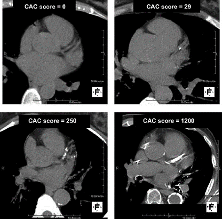
CAC scan
- Main question
- How much calcified plaque is in the coronary arteries?
- Best use
- Refining future heart risk when the result could change a prevention decision.
- What to know
- A zero score does not exclude noncalcified plaque. CAC does not show whether a narrowing limits blood flow.
- Radiation and contrast
- CT radiation; usually no injected contrast.
CCTA
- Main question
- Where are plaque and narrowing in the coronary arteries?
- Best use
- Evaluating suspected coronary disease, including selected people with stable chest symptoms.
- What to know
- Shows calcified and noncalcified plaque. A narrowing may need a separate blood-flow assessment; heavy calcium or poor image quality can limit interpretation.
- Radiation and contrast
- CT radiation and iodinated IV contrast.
Stress testing
- Main question
- Does exercise or medication-induced stress reveal inadequate blood supply to the heart muscle?
- Best use
- Evaluating suspected flow-limiting disease, symptoms or exercise capacity.
- What to know
- A normal stress test does not mean there is no plaque. The different stress tests below answer this question in different ways.
- Radiation and contrast
- Depends on the test: ECG, echo and MRI use no ionizing radiation; SPECT and PET use radioactive tracers.
What Heartflow and Cleerly add to CCTA
These services analyze the CCTA images. Plaque analysis measures the disease in the artery wall; ischemia analysis estimates whether disease is limiting blood supply. The software analysis itself adds no radiation or contrast to an adequate existing scan. The underlying CCTA still uses both. [8] [12]
Heartflow
- Plaque Analysis + Plaque Staging
- Measures plaque amount and composition, then summarizes the burden by stage.
- FFRCT Analysis
- Uses a computer model of coronary blood flow to estimate the effect of specific narrowings.
- Roadmap Analysis
- Maps coronary anatomy and the location and severity of narrowing.
PCI Navigator is a related tool for planning a coronary stent procedure.
Cleerly
- Plaque Analysis
- Measures plaque amount, type and location, together with artery narrowing.
- Cleerly ISCHEMIA
- Uses AI analysis of plaque and artery features to estimate the likelihood of reduced blood supply in each vessel.
- Cleerly COMPARE
- Compares plaque findings between CCTA scans obtained at different times.
A new follow-up CCTA is a new scan, with its own radiation and contrast exposure.
Compare the stress tests
Exercise ECG
- How it works
- A treadmill or bicycle test records the ECG, symptoms, blood pressure and exercise capacity.
- Radiation
- None. No imaging or radioactive tracer.
- Main tradeoff
- A useful option when you can exercise and your resting ECG can be interpreted for ischemia. It is less diagnostically accurate than stress imaging.
Stress echocardiogram
- How it works
- Ultrasound compares heart-muscle contraction at rest and with exercise or medication stress.
- Radiation
- None. An ultrasound contrast agent may improve unclear images; it is not radioactive.
- Main tradeoff
- Good diagnostic accuracy without radiation. Poor ultrasound windows or inadequate stress can reduce sensitivity.
Nuclear SPECT
- How it works
- A radioactive tracer maps relative blood supply to the heart muscle at rest and after exercise or medication stress.
- Radiation
- Yes, from the tracer; dose depends on the protocol.
- Main tradeoff
- Widely used, but image artifacts or similarly reduced flow across several arteries can obscure disease.
Nuclear PET
- How it works
- A radioactive tracer maps blood supply; PET can also quantify blood flow and flow reserve.
- Radiation
- Yes. PET is still a nuclear test.
- Main tradeoff
- When nuclear imaging is chosen, guidelines favor PET over SPECT when available because of better diagnostic accuracy and fewer inconclusive studies.
Stress MRI / CMR
- How it works
- Usually uses medication stress and gadolinium contrast to image heart-muscle blood supply; it also assesses function and scar.
- Radiation
- None. MRI uses magnetic fields and radio waves.
- Main tradeoff
- Detailed imaging without radiation. Kidney function, implanted-device compatibility, enclosed-space tolerance and local availability affect suitability.
Three questions to ask
- Are we measuring plaque, explaining symptoms or checking blood flow?
- What would the result change in my care?
- If a CT add-on or a second test is proposed, what unanswered question will it resolve?
New or severe chest pressure, shortness of breath or fainting needs urgent assessment rather than an elective screening appointment.
Keep learning
Understanding CAC scans · Heart Health & Screening
Sources
- ACC: imaging choices in the multisociety chest-pain guideline.
- SCCT coronary CT angiography consensus.
- ASE stress echocardiography guideline.
- American Heart Association: exercise stress testing.
- American Heart Association: PET and SPECT.
- American Heart Association: cardiac MRI.
- SCMR standardized CMR protocols.
- Heartflow: current product platform and patient information.
- Heartflow: Plaque Analysis, FFRCT Analysis and Roadmap Analysis.
- Cleerly: Plaque Analysis and ISCHEMIA.
- Cleerly COMPARE.
- Cleerly patient FAQs.