
Lower-back pain can be intense, frightening and disabling. Yet the severity of the pain does not always tell us how much structural damage is present—or whether a scan will reveal the reason it hurts.
That apparent contradiction is one of the most important ideas in back-pain medicine. Some people have dramatic-looking disc degeneration on an MRI and feel no pain. Others have severe pain with no single abnormality that explains it. Pain is real, but it is produced by a living system: joints, discs, muscles, nerves, immune signals, the brain, sleep, stress, movement, previous injury and metabolic health can all contribute.
Most new episodes improve substantially with time and appropriate movement. A smaller group follows a persistent or recurring course. An even smaller group has a serious underlying condition that changes the evaluation urgently. The goal is not to scan everyone or dismiss everyone. It is to recognize the pattern, look for meaningful warning signs and use testing when the result can change what happens next.
Lower-back pain is a symptom, not one diagnosis
The majority of lower-back pain seen in primary care is called nonspecific low-back pain. This does not mean that the pain is vague, psychological or unimportant. It means that no single fracture, infection, tumor, compressed nerve or inflammatory disease accounts for the entire presentation.
Other recognizable patterns include:
- Radicular pain or sciatica: irritation of a spinal nerve root can produce pain traveling into the buttock or leg, sometimes with numbness, tingling or weakness.
- Spinal stenosis: narrowing around spinal nerves can cause leg pain, heaviness or weakness during standing and walking, often improving with sitting or leaning forward.
- Vertebral compression fracture: more likely after significant trauma or in people with osteoporosis, advanced age or prolonged glucocorticoid exposure.
- Inflammatory back pain: often begins at a younger age and may involve prolonged morning stiffness, night pain and improvement with movement. Axial spondyloarthritis is one important cause.
- Referred pain: pain arising from the abdomen, pelvis, kidneys, blood vessels or other structures can sometimes be felt in the back.
These categories can overlap. A disc can irritate a nerve without being the only reason pain persists, and a person can have age-related spinal changes plus muscular, sleep-related or metabolic factors that affect recovery.
Severe pain does not necessarily mean dangerous disease
Serious spinal pathology is uncommon in routine primary care. In a prospective cohort of 1,172 people with acute lower-back pain, 11—0.9%—were ultimately found to have a serious cause, most commonly vertebral fracture.[11]
That study also revealed why a checklist alone is not enough: 80.4% of participants had at least one commonly cited red flag. A single finding can be nonspecific. The diagnostic value comes from the combination of the history, examination, risk profile and evolution of symptoms.
Features that can meaningfully change the level of concern include:
- new urinary retention, loss of bowel control or numbness in the saddle area;
- new or progressive leg weakness or a major neurologic deficit;
- fever or systemic illness with risk factors for spinal infection;
- significant trauma, especially when bone strength may be reduced;
- a relevant current or previous cancer together with new unexplained back pain;
- unexplained weight loss or pain that is persistently progressive and does not behave like ordinary mechanical pain.
These findings do not automatically prove that a dangerous condition is present. They signal that the usual watchful, movement-based approach may not be sufficient and that prompt clinical assessment may be appropriate.
Why an MRI can be both useful and misleading
Imaging is powerful when it answers a focused question. It is much less useful when it is treated as a fishing expedition.
A systematic review of 33 studies involving 3,110 people without back pain found that degenerative findings became increasingly common with age.[12] Disc degeneration appeared in an estimated 37% of asymptomatic 20-year-olds and 96% of asymptomatic 80-year-olds. Disc bulges increased from 30% to 84% across the same age range.
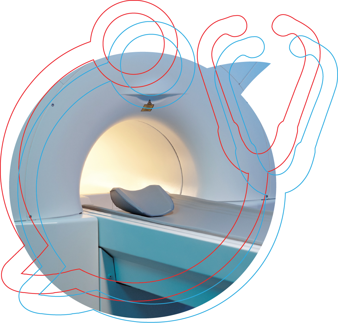
These findings are not meaningless. Some imaging abnormalities are more common in people with pain, and a scan can identify important pathology. But an MRI report is not a pain meter. A disc bulge may be central to one person's symptoms and incidental in another.
This is why the scan must be interpreted alongside the clinical pattern:
- MRI is especially useful when clinicians are concerned about compressed neural structures, infection, cancer, cauda equina syndrome or persistent and progressive symptoms for which an invasive intervention is being considered.
- CT is valuable for detailed evaluation of bone and suspected fractures and can be used when MRI is unavailable or cannot be performed.
- Plain radiographs can answer selected questions about fracture, alignment or structural disease but have limited value for uncomplicated nonspecific pain.
Randomized studies of routine early imaging have generally not shown meaningful improvements in pain or physical function for people without indicators of serious disease.[13,14] The question is therefore not simply, “Can we obtain an image?” It is, “What clinical question will this image answer, and will the answer alter management?”
Recovery is usually favorable—but not always linear
Many community episodes resolve rapidly. In a 2023 community inception cohort of 366 new episodes, the median duration was five days. Recovery occurred in 70% before one week, 86.1% before three weeks and 93.5% before 12 weeks.[15]
People presenting for clinical care can have a different course. Some recover quickly, some improve gradually, some fluctuate and some develop persistent symptoms. Previous episodes, longer symptom duration and greater initial pain or disability can influence the trajectory.[16]
Recurrence is also common. A person can recover completely from one episode and experience another months later. That does not necessarily mean the spine is progressively deteriorating. It means that recovery and prevention deserve separate attention.
Movement is usually part of the solution
During a painful episode, movement may feel threatening. Prolonged bed rest, however, can reduce confidence and conditioning without accelerating recovery. For most nonspecific back pain, the better strategy is to remain as active as symptoms reasonably allow and gradually rebuild capacity.
Useful components can include:
- walking;
- graded return to normal daily activity;
- trunk endurance and motor-control work;
- hip and lower-extremity strength;
- physical therapy when pain, weakness, fear or recurrent episodes make self-directed progress difficult;
- heat for short-term comfort during an acute flare.
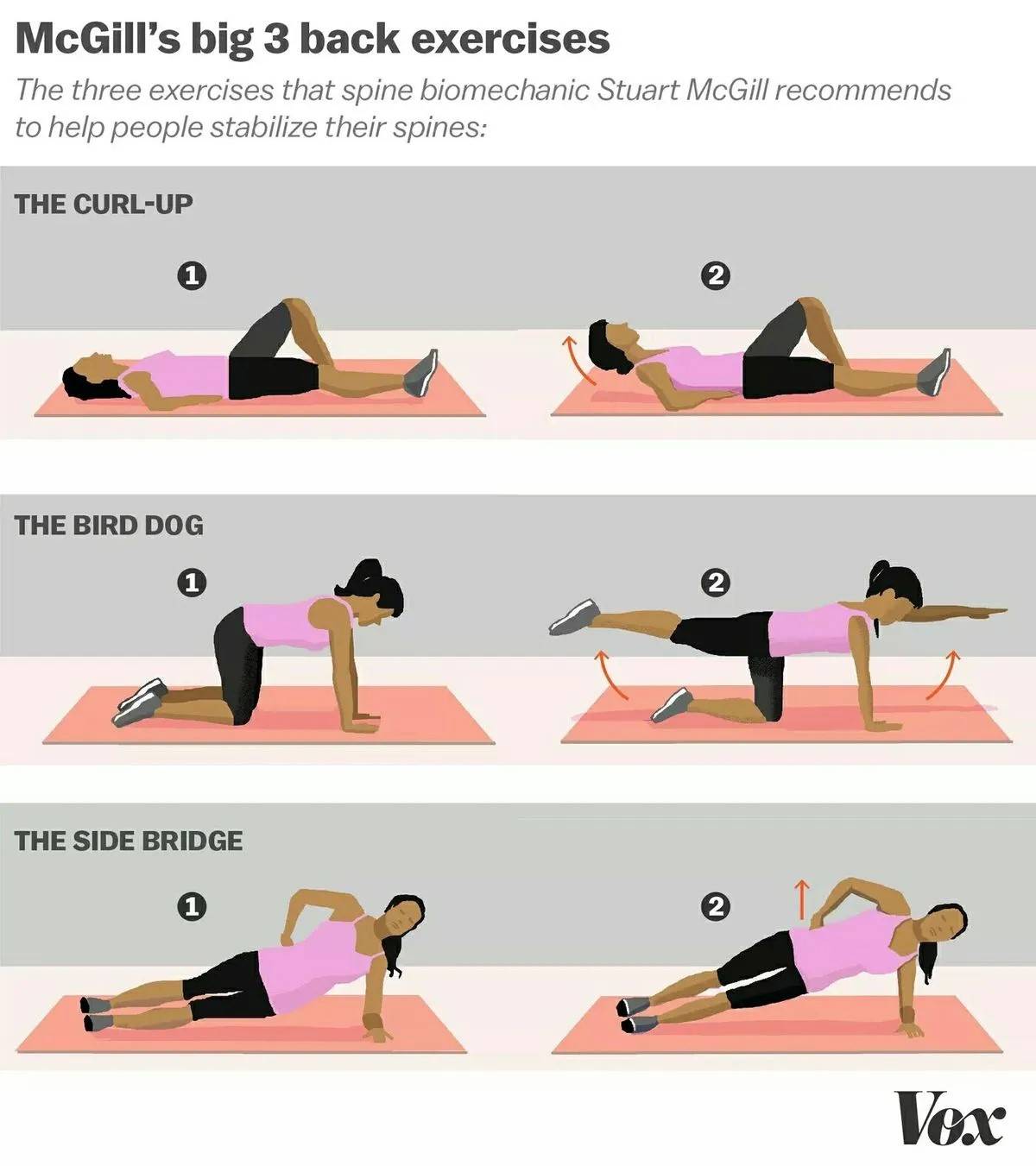
The McGill Big Three—the modified curl-up, side plank and bird dog—are familiar trunk-endurance exercises and can be useful within a broader program. They are tools, not a universally superior protocol. Exercise selection should reflect the person's symptoms, capabilities, goals and response.
Walking deserves particular attention because it is accessible and scalable. In the 2024 WalkBack randomized trial, 701 adults who had recently recovered from nonspecific lower-back pain received either an individualized progressive walking-and-education program or no intervention.[17] The program reduced the risk of an activity-limiting recurrence. Median time to recurrence was 208 days with the intervention versus 112 days in the control group.
That result supports a practical message: rehabilitation does not always require a complicated or highly specialized routine. A progressive plan that people can actually continue may meaningfully change recurrence.
Core, glute and lower-back stability
This progression combines the McGill Big Three with glute strengthening and hip mobility. Start with controlled, comfortable movements, then gradually increase the challenge as your strength and confidence improve.
- Foundation: Modified curl-ups, bird dogs and bent-knee side planks; bodyweight glute bridges and dead bugs; 90/90 hip stretches, kneeling hip-flexor stretches and assisted Cossack squats.
- Build control: Progress to side planks with feet stacked or the top foot forward, small-circle stir-the-pot, planks with slow mountain climbers, and kneeling or standing Pallof presses. Add bodyweight bench hip thrusts, unassisted Cossack squats, wall quad/hip-flexor stretches and seated center-split stretches.
- Add strength and endurance: Progress to larger stir-the-pot circles, side planks with a reach or weight, mountain climbers with a pause, and Pallof presses with an overhead extension. Add dumbbell or barbell hip thrusts, Cossack squats with a reach or weight, banded hip-flexor stretches and supported wall or sliding center-split stretches.
Optional glute work: Frog pumps, banded glute-bridge marches and clamshells.
Choose exercises that suit your symptoms and ability; a physical therapist can help. Progress at your own pace, avoid forcing stretches, and stop any exercise that worsens symptoms.
What medication studies add to the picture
Medication can be one part of short-term symptom management, but different drug classes do not have equal evidence and the effect often depends on whether pain is nonspecific, radicular or caused by another condition.
Consider the PACE trial, a double-blind randomized study of 1,652 people with acute lower-back pain.[18] Median recovery time was 17 days with regularly scheduled acetaminophen, 17 days with acetaminophen taken as needed and 16 days with placebo. Acetaminophen did not accelerate recovery or improve the measured secondary outcomes in that trial.
NSAIDs and short courses of some skeletal muscle relaxants can provide symptom relief for selected patients, although gastrointestinal, kidney, cardiovascular, sedation and interaction risks matter when choosing them.[1]
Systemic corticosteroids require more nuance than a simple effective-or-ineffective label. In a randomized trial of 269 people with acute radicular pain and MRI-confirmed disc herniation, a short course of oral prednisone produced modest improvement in function but did not meaningfully improve pain compared with placebo.[19]
Treatment should therefore follow the pain pattern and the person—not simply the intensity of the pain.
Why lower-back pain also belongs in metabolic health
Lower-back pain is not always an inflammatory disorder, and metabolic dysfunction is not the explanation for every painful back. Still, the connection is strong enough that it should not be treated as an afterthought.
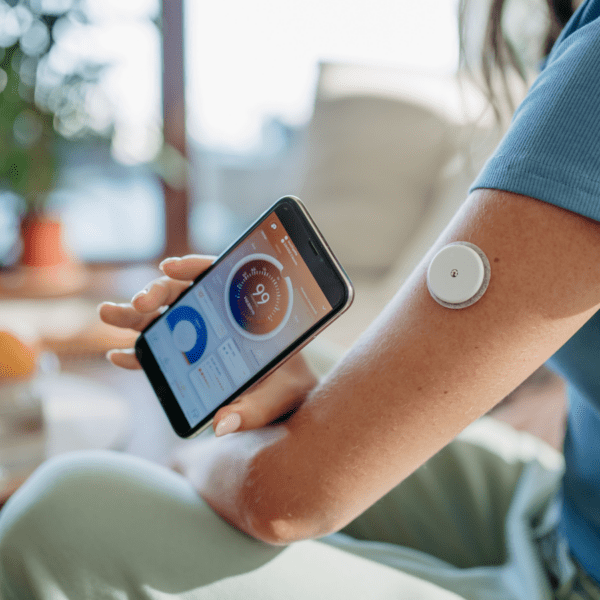
Body composition can affect the spine mechanically through load and movement. It can also affect muscle quality, physical capacity, sleep, vascular health, immune signaling and pain sensitivity. Sedentary behavior can sit in the middle of the relationship: pain reduces movement, while reduced movement can worsen conditioning and metabolic health.
A large Mendelian-randomization study used genetic variants associated with BMI to examine more than 450,000 participants.[20] A one-standard-deviation increase in BMI—approximately 4.65 kg/m²—was associated with 15% higher odds of back pain and 20% higher odds of chronic back pain. Mendelian randomization cannot resolve every mechanism, but it strengthens the argument that the relationship is not explained entirely by reverse causation.
Newer work has examined metabolic syndrome more directly. A 2026 analysis combined cross-sectional NHANES data with genetic causal-inference methods.[21] Metabolic syndrome was associated with lower-back pain across three commonly used definitions. The genetic analysis particularly implicated waist circumference and hypertension. Interestingly, C-reactive protein did not mediate the observed association.
That last finding is important. The metabolic connection should not be reduced to “inflammation causes pain.” Abdominal adiposity, blood pressure, vascular function, muscle capacity, movement and nervous-system biology may contribute through several interacting pathways. The relationship is biologically richer than one laboratory marker.
Our companion article, Chronic Pain and Metabolic Health, explores the emerging intervention literature on low-carbohydrate and ketogenic nutrition, inflammatory markers and several chronic-pain conditions. Keeping that research in its own article allows this page to remain focused on lower-back pain while giving interested readers a clear next step.
A practical framework for understanding an episode
When lower-back pain appears, the most useful questions are often:
- Is this a new, recurrent or persistent episode?
- Is the pain confined to the back, or does it travel into the leg?
- Are strength, sensation, walking or bowel and bladder function changing?
- Was there trauma, systemic illness or another reason to suspect a specific cause?
- What movements aggravate or relieve the symptoms?
- Are sleep, stress, inactivity or metabolic health making recovery harder?
- Would imaging change the plan—or merely give a name to an age-related finding?
This framework replaces two unhelpful extremes: assuming every painful back is damaged, or assuming all back pain should simply be ignored.
The practical takeaway
Most lower-back pain is not caused by a dangerous disease, but the pain can still be severe and disruptive. The best evaluation combines the history, examination and evolution of symptoms rather than relying on one red flag or one scan.
Imaging is most valuable when it answers a clinical question that can change management. Movement and progressive rehabilitation are central to recovery and prevention for many people. Medications can be useful selectively, but they do not replace an accurate understanding of the pain pattern.
Metabolic health belongs in the conversation as well. Body composition, blood pressure, physical capacity, sleep, inflammation and nervous-system function can interact with pain—especially when it becomes chronic or recurrent. Lower-back pain is therefore appropriately housed within Metabolic Health → Chronic Pain & Inflammation, while also appearing visually in Exercise, Strength & Mobility.
References
Source references
-
Qaseem A, et al. (2017). Noninvasive Treatments for Acute, Subacute, and Chronic Low Back Pain: A Clinical Practice Guideline From the American College of Physicians. Ann Intern Med. 166:514–530.
-
Chou R, et al. (2007). Diagnosis and Treatment of Low Back Pain: A Joint Clinical Practice Guideline from the American College of Physicians and the American Pain Society. Ann Intern Med. 147(7):478–491.
-
Chou R, et al. (2011). Diagnostic Imaging for Low Back Pain: Advice for High-Value Health Care From the American College of Physicians. Ann Intern Med. 154:181–189.
-
Patel ND, et al. (2016). ACR Appropriateness Criteria Low Back Pain. J Am Coll Radiol. 13(9):1069–1078.
-
Wheeler SG, et al. (2017). Evaluation of low back pain in adults. UpToDate.
-
Knight CL, et al. (2017). Treatment of acute low back pain. UpToDate.
-
Chou R, et al. (2017). Subacute and chronic low back pain: Nonpharmacologic and pharmacologic treatment. UpToDate.
-
Hsu PS, et al. (2017). Acute lumbosacral radiculopathy: Pathophysiology, clinical features, and diagnosis. UpToDate.
-
ACEP Choosing Wisely Recommendation. Retrieved from Choosing Wisely.
-
AAFP Choosing Wisely Recommendation. Retrieved from AAFP.
Additional evidence
-
Henschke N, Maher CG, Refshauge KM, et al. Prevalence of and screening for serious spinal pathology in patients presenting to primary care settings with acute low back pain. Arthritis Rheum. 2009;60(10):3072–3080. doi: 10.1002/art.24853. PubMed
-
Brinjikji W, Luetmer PH, Comstock B, et al. Systematic literature review of imaging features of spinal degeneration in asymptomatic populations. AJNR Am J Neuroradiol. 2015;36(4):811–816. doi: 10.3174/ajnr.A4173. PubMed
-
Kendrick D, Fielding K, Bentley E, et al. Radiography of the lumbar spine in primary care patients with low back pain: randomised controlled trial. BMJ. 2001;322:400–405. doi: 10.1136/bmj.322.7283.400. PubMed
-
Gilbert FJ, Grant AM, Gillan MGC, et al. Low back pain: influence of early MR imaging or CT on treatment and outcome—multicenter randomized trial. Radiology. 2004;231(2):343–351. doi: 10.1148/radiol.2312030886. PubMed
-
de Campos TF, et al. Prognosis of a new episode of low-back pain in a community inception cohort. Eur J Pain. 2023. PubMed
-
Kongsted A, Kent P, Axen I, Downie AS, Dunn KM. Trajectories of acute low back pain: a latent class growth analysis. Pain. 2016. PubMed
-
Pocovi NC, et al. Effectiveness and cost-effectiveness of an individualised, progressive walking and education intervention for the prevention of low back pain recurrence in Australia (WalkBack): a randomised controlled trial. Lancet. 2024. PubMed
-
Williams CM, Maher CG, Latimer J, et al. Efficacy of paracetamol for acute low-back pain: a double-blind, randomised controlled trial. Lancet. 2014;384(9954):1586–1596. doi: 10.1016/S0140-6736(14)60805-9. PubMed
-
Goldberg H, Firtch W, Tyburski M, et al. Oral steroids for acute radiculopathy due to a herniated lumbar disk: a randomized clinical trial. JAMA. 2015;313(19):1915–1923. doi: 10.1001/jama.2015.4468. PubMed
-
Elgaeva EE, Tsepilov Y, Freidin MB, et al. Examining causal effects of body mass index on back pain: a Mendelian randomization study. Eur Spine J. 2020;29:686–691. doi: 10.1007/s00586-019-06224-6. PubMed
-
Wang et al. Metabolic Syndrome and Low Back Pain: Evidence from Cross-Sectional and Mendelian Randomization Analysis. 2026. PubMed